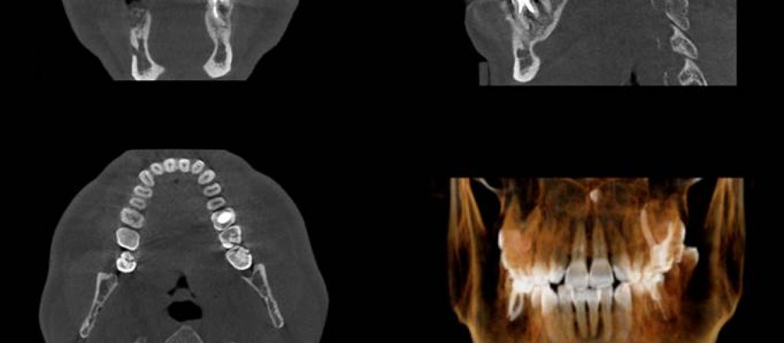
A Cone Beam CT scanner uses a special technology to generate 3D images of dental structures, soft tissues, nerve paths, and bone in the craniofacial region in a single scan. At Oral & Maxillofacial Surgery of Carbondale, CBCT imaging is indicated for deeply impacted teeth, implants, some bone grafting procedures, pathology such as cysts, tori removal, supernumerary teeth, and evaluation of the jaw, sinuses, nerve canals, and nasal cavity.
Images obtained with a CBCT allow for more precise treatment planning and predictable surgical results. We take our own CBCT imaging to ensure accurate data collection. There are many CBCT machines on the market and the imaging and quality you get from one machine to another can vary widely. Different machines have varying field of view (FOV), voxel size, and visualization software. A Kodak 9000 3D has a max FOV of only 4x5 cm, which would be adequate for endo to visualize 1 or 2 teeth and many general dentists have a machine with max FOV of 8x8 or 8x10 cm, which are all far too small for oral surgery. Voxel sizes for CBCT machines vary and some machines have multiple voxel sizes. If the voxel size is larger, the image resolution decreases and that affects the diagnostic quality.
CBCT image quality and accuracy can also be affected if your CBCT is not calibrated regularly. Other factors that affect image quality are artifacts such as when jewelry is not removed prior to taking imaging or metal artifacts due to existing dental implants, crowns, and bridges. We utilize metal artifact reduction during reconstruction of the CBCT imaging to best plan the surgery. Also, patient motion can cause artifacts and result in bad data. If you are seen as patient at our office and a CBCT is needed, we will take our own imaging to ensure we provide the highest level of surgical care and minimize any associated risks.

